Mostrar el registro sencillo del ítem
Quantification of Callose Deposition in Plant Leaves
| dc.contributor.author | Scalschi, Loredana | |
| dc.contributor.author | Llorens, Eugenio | |
| dc.contributor.author | Camañes, Gemma | |
| dc.contributor.author | Pastor, Victoria | |
| dc.contributor.author | Fernández Crespo, Emma | |
| dc.contributor.author | Flors, Victor | |
| dc.contributor.author | García Agustín, Pilar | |
| dc.contributor.author | Vicedo, Begonya | |
| dc.date.accessioned | 2016-02-22T10:33:51Z | |
| dc.date.available | 2016-02-22T10:33:51Z | |
| dc.date.issued | 2015-10 | |
| dc.identifier.citation | SCALSCHI, Loredana, et al. Quantification of Callose Deposition in Plant Leaves. 2015. | ca_CA |
| dc.identifier.issn | 2331-8325 | |
| dc.identifier.uri | http://hdl.handle.net/10234/150985 | |
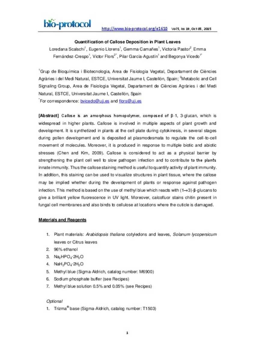
| dc.description.abstract | Callose is an amorphous homopolymer, composed of β-1, 3-glucan, which is widespread in higher plants. Callose is involved in multiple aspects of plant growth and development. It is synthetized in plants at the cell plate during cytokinesis, in several stages during pollen development and is deposited at plasmodesmata to regulate the cell-to-cell movement of molecules. Moreover, it is produced in response to multiple biotic and abiotic stresses (Chen and Kim, 2009). Callose is considered to act as a physical barrier by strengthening the plant cell well to slow pathogen infection and to contribute to the plant’s innate immunity. Thus the callose staining method is useful to quantify activity of plant immunity. In addition, this staining can be used to visualize structures in plant tissue, where the callose may be implied whether during the development of plants or response against pathogen infection. This method is based on the use of methyl blue which reacts with (13)--glucans to give a brilliant yellow fluorescence in UV light. Moreover, calcofluor stains chitin present in fungal cell membranes and also binds to cellulose at locations where the cuticle is damaged. | ca_CA |
| dc.description.sponsorShip | Authors thank Universitat Jaume I and the National R&D Plan (AGL2010-22300-C03-02, Spain for funding support. | ca_CA |
| dc.format.extent | 6 p. | ca_CA |
| dc.format.mimetype | application/pdf | ca_CA |
| dc.language.iso | eng | ca_CA |
| dc.publisher | Bio-protocol LLC | ca_CA |
| dc.relation.isPartOf | Bio-protocol 5(19), 2015 | ca_CA |
| dc.rights | © 2015 Bio-protocol LLC. | ca_CA |
| dc.rights.uri | http://rightsstatements.org/vocab/InC/1.0/ | * |
| dc.title | Quantification of Callose Deposition in Plant Leaves | ca_CA |
| dc.type | info:eu-repo/semantics/article | ca_CA |
| dc.rights.accessRights | info:eu-repo/semantics/openAccess | ca_CA |
| dc.relation.publisherVersion | http://www.bio-protocol.org/e1610 | ca_CA |
Ficheros en el ítem
Este ítem aparece en la(s) siguiente(s) colección(ones)
-
CAMN_Articles [566]


